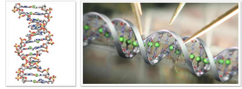
Conducting DNA nanowire/nanodevice development
DNA is one of the most promising one-dimensional nanomaterials because of its adjustable length and self-assembly properties. It may be useful as a one-dimensional nanowire or nanodevice in electronic applications; however, it is of great interest to reveal the electrical conduction mechanism of DNA. The conductivity of DNA increases dramatically when it is doped with Ni ions to form Ni-DNA (M-DNA). Our studies demonstrate that Ni-DNA is a conducting nanowire. The the charges in Ni2+-doped DNA are transported through the Ni2+-mediated p-p stacking corridor.
Native bio-nanoparticle drug delivery system development
For hydrophobic drug delivery, it is important to consider the physicochemical properties of macromolecules, cell uptake, and the usage of the delivery system. A good hydrophobic drug delivery system (DDS) with native-like physicochemical properties and good cell permeability is highly desired. Modified natural transporter/protein would be an excellent candidate for DDS.
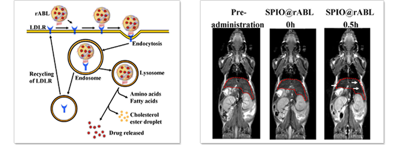
Low-density lipoprotein (LDL) is a native nanoparticle and its size is approximately 18–25 nm. Plasma LDL can be cleared from circulation through LDL receptor (LDLR)-mediated endocytosis. This allows for the transfer of LDL into endosomes, where the pH drops, causing LDL to dissociate from the receptor. The receptor is then recycled to the surface of the cell, while LDL is transferred into the lysosomes for degradation. This pathway could be useful for drug delivery, and may then be directed toward tumors expressing LDLR. It has been demonstrated that LDLR is overexpressed in various human cancer cell lines. A LDL-based hydrophobic/amphiphilic DDS can be internalized and degraded by regular cell metabolism, and these properties can be used in targeted therapies for cancer. In our previous study indicated that the reconstituted apoB lipoparticle (rABL) is an ideal carrier for transporting hydrophobic and amphiphilic compounds.
Ni-DNA nanowire device
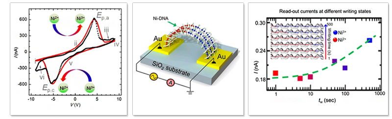
DNA is an one-dimensional nanomaterials with the properties of self-assembly and length controllable by bio-synthesis. The electrical property of Ni-DNA has been characterized via AFM, demonstrating that Ni-DNA acts as a conducting nanowire. It has also been demonstrated that Ni-DNA acts like a conducting wire with an insulated shell to prevent intermolecular electron leakage. Furthermore, the device with the Ni2+-chelated DNA (Ni-DNA) connecting two gold electrodes, exhibited a negative differential resistance (NDR) behavior, when the measurement of current response in a cyclic-voltage scan (I-V) was performed under an ambient and water-free condition. Each Ni ion in Ni-DNA exhibits low (Ni2+) or high (Ni3+) oxidation state and can be switched sequentially by applying bias voltage with different polarities and writing times. The ratio of low and high oxidation states of Ni ions in Ni-DNA represents a programmable multi-state memory system with an added capacitive component, in which multi-state information can be written, read, and erased. This study also indicates the biomolecules-based self-organized nanostructure can be used as a template for nanodevices fabrication.
Ultra-sensitive biosensor platform development
1. Rapid and highly sensitive detection of Enterovirus 71 by using nanogoldenhanced electrochemical impedance spectroscopy
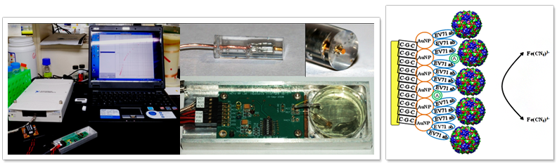
Enterovirus 71 (EV71) infection is an emerging infectious disease causing neurological complications and/or death within two to three days after the development of fever and rash. A low viral titre in clinical specimens makes the detection of EV71 difficult. Conventional approaches for detecting EV71 are time consuming, poorly sensitive, or complicated, and cannot be used effectively for clinical diagnosis. Furthermore, EV71 and Coxsackie virus A16 (CA16) may cross react in conventional assays. Therefore, a rapid, highly sensitive, specific, and user-friendly test is needed. We developed an EV71-specific nanogold-modified working electrode for electrochemical impedance spectroscopy in the detection of EV71. Our results show that EV71 can be distinguished from CA16, Herpes simplex virus, and lysozyme, with the modified nanogold electrode being able to detect EV71 in concentrations as low as 1 copy number/50 ml reaction volume, and the duration between sample preparation and detection being 11 min. This detection platform may have the potential for use in point-of-care diagnostics.
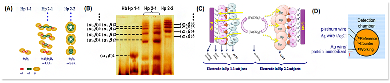
2. Human heptoglobin typing and concentration determination
Haptoglobin (Hp) is an acute phase protein that binds free hemoglobin (Hb), preventing Hb-induced oxidative damage in the vascular system. There are three phenotypes in Human Hp, which heterogeneous polymorphic structures and varying concentrations in plasma have been attributed to the cause of diseases, and outcome of clinical treatments. Different phenotypes of Hp may compose of the same subunits but different copy numbers, rendering their determination difficult by a single procedure. In this study, we have developed a simple, fast, reliable, and sensitive method, using label-free nanogold modified bioprobes coupled with self-development electrochemical impedance spectroscopy (EIS). By this method, probe surface charge transfer resistance is detected. The relative charge transfer resistance ratios for Hp 1-1, Hp 2-1, and Hp 2-2 were characterized. We were able to determine protein size difference within 3 nm, and the linear region of the calibration curve for Hp levels in the range of 90 pg/ml and 90 mg/ml (~1fM to 1pM). We surmise that similar approaches can be used to investigate protein polymorphism and altered protein-protein interaction associated with diseases.
- Ultra high sensitive biosensing platform have been developed in my lab.
- This platform can be used to detect infectious, emerging disease on site.
- This platform can be used to detect and quantify the proteins and chemicals.
- This platform can be used for detecting intermolecular interactions.
http://nanotechweb.org/cws/article/lab/46448

Protein folding and mechanism study
Protein is a macromolecule which posses self-organization and self-assembly properties. Protein folds spontaneously in the heterogeneous environment. However, some of them may aggregate with each other during the folding process. Therefore, both folded and aggregated protein molecules can be observed under certain folding condition. These two states, (unfolded/folded), coexist phenomenon indicates that the reaction of protein folding is a “first-order like phase transition” process. By combing experimental approaches and molecular simulation analysis, we can conclude that the protein aggregation process is a competitive process between “spontaneous folding” and “diffusion limited aggregation” reactions. Meanwhile, the protein self-stabilization time can be revealed. Moreover, by manipulating the folding process, room-temperature protein base molecular magnet can be synthesized. The protein selfassembly nano-pattern can be performed on silicon or metal substrate.
Single molecular studies
Tight-Turn Paperclip DNA Triplex in Solution
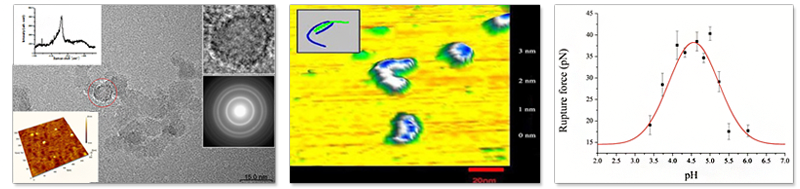
DNA triplex modulates gene expression by forming stable conformation in physiological condition. However, it is not feasible to observe this unique molecular structure of large molecule with 54 oligodeoxynucleotides directly by conventional nuclear magnetic approach. In this study, we observed directly single molecular images of paperclip DNA triplexes formation in a buffer solution of pH 6.0 by atomic force microscopy (AFM). Meanwhile, a diffuse “tail” of unwound DNAwas observed in pH8.0 solution. This designable approach in visualizing the overall structures and shapes of oligo-DNAs at the single molecular level, by AFM, is applicable to other biopolymers as well.
Nanodiamonds
Nanodiamond ND is surrounded by layers of graphite on its surface. This unique structure feature creates unusual fluorescence spectra, which can be used as an indicator to monitor its surface modification. Meanwhile, the impurity, nitroso CuNvO inside the ND can be photolyzed by two-photon absorption, releasing NO to facilitate the formation of a sp3 diamond structure in the core of ND and transforming it into a sp2 graphite structure. Such a conformational transition enlarges the size of ND from 8 to 90 nm, resulting in a popcornlike structure. This transition reaction may be useful as nanoknives in biomedical application. Applied Physics Letters. 93,033905
Bio-photonics and mechanics
Triplex DNA formation/force analysis
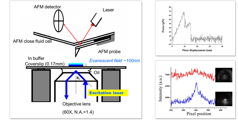
We have detected unambiguously the dynamics of 17-mer DNA triplex dissociation mechanism in molecular level. Technique of fluorescence resonance energy transfer (FRET) has been used as an indicator for inter-molecular interaction in nanometer range, and atomic force microscopy (AFM) is employed to address the single molecule with sub-angstrom precision. By combining these two techniques, the triplex rupturing event is ascertained to involve one or a few triplex DNA molecule only. Meanwhile, the maximum DNA triplex rupture force is found between the pH of 4.12 and 5.00. This molecular mechanism is consistent with the macroscopic observation of NMR studies. These results indicated that the FRET-combining AFM detection system can be used for revealing molecular interaction in nano-meter scale.
Functional bio-nanomaterials development

Targeting nanoblast for cancer therapy by functionalized Nanodiamonds
It is highly desire to develop a therapeutic, observable nanoparticle complex for specifically targeting in cancer therapy. Growth hormone (GH) and its antagonists have been explored as cancer cell targeting molecules for both imaging and therapeutic applications. In this study, a low toxic, biocompatible, therapeutic, and observable GH-nanoparticle complex for specifically targeting growth hormone receptor (GHR) in cancer cells, has been synthesized by conjugating GH with green fluorescence protein and carboxylated nanodiamond. Moreover, we have shown that this complex can be triggered by laser irradiation to create a “nanoblast” and induce cell death in the non-small lung cancer cell line A549 via the apoptotic pathway. This laser-mediated cancer-targeting platform can be wildly used in cancer therapy.

Mechanism for ND nanoblast
Nanodiamond (ND) is surrounded by layers of graphite on its surface. This unique structure feature creates unusual fluorescence spectra, which can be used as an indicator to monitor its surface modification. Meanwhile, the impurity, nitroso (C-N=O) inside the ND can be photolyzed by two-photon absorption, releasing NO to facilitate the formation of a sp3 diamond structure in the core of ND and transforming it into a sp2 graphite structure. Such a conformational transition enlarges the size of ND from 8 to 90 nm, resulting in a popcornlike structure. This transition reaction may be useful as nanoknives in biomedical application.
Biomaterials synthesis and characterization
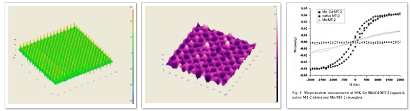
First room temperature ferromagnetic protein in the world !!
The paper reports the methods of preparing molecular magnets and patterning of the molecules on a semiconductor surface. A highly magnetically aligned metallothionein containing Mn and Cd (Mn,Cd-MT-2) is first synthesized, and the molecules are then placed into nanopores prepared on silicon (0 0 1) surfaces using electron beam lithography and reactive ion-etching techniques. We have observed the self-assemble growth of the MT molecules on the patterned Si surface such that the MT molecules have grown into rod or ring type three dimensional nanostructures, depending on the patterned nanostructures on the surface. We also provide scanning electron microscopy, atomic force microscopy, and magnetic force microscope studies of the molecular nanostructures. This engineered molecule shows molecular magnetization and is biocompatible with conventional semiconductors. These features make Mn,Cd-MT-2 a good candidate for biological applications and sensing sources of new nanodevices. Using molecular self-assembly and topographical patterning of the semiconductor substrate, we can close the gap between bio-molecules and nanoelectronics built into the semiconductor chip.
Biomaterials
Volume 28, Issue 11, April 2007, Pages 1941–1947
開發鈀奈米薄膜電化學感測平台用於病毒檢測與抑制分子篩選
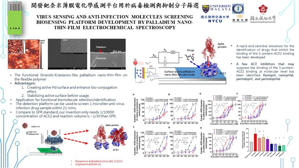
Virus sensing and anti-infection molecules screening biosensing platform development by palladium nano-thin-film electrochemical spectroscopy
●技術簡介:
新型冠狀病毒藉由與人類細胞的ACE2受體結合感染細胞。本技術以新穎鈀薄膜電化學阻抗偵測系統為基礎,研發出新型冠狀病毒偵測平台並可用於篩選抑制新型冠狀病毒感染的藥物,只需要取樣1微升(μL)的樣品,就能在21分鐘內偵測辨識新型冠狀病毒或篩選能影響病毒與ACE2結合的藥物。
●技術之科學突破性:
傳統病毒偵測常以PCR或ELISA方法,本技術採用本團隊所開發之鈀奈米薄膜電極電化學系統,建立病毒棘蛋白與ACE2交互作用之感測平台。以偵測病毒棘蛋白與ACE2交互作用為模型乃因能提供病毒感染之功能性測試,在學理上可比較不同病毒變異株對人類ACE2結合能力之差異,並推測變種病毒對人體感染之差異性以提供其分子作用機制的探討。此成果發表於今年mSphere期刊。此外,本偵測系統亦可用於感測中和抗體之等效滴度,此乃傳統PCR或ELISA檢測方法所無法或難以達到的精確度。由於偵測的分子交互作用與病毒感染力高度關連,因此可用以篩選抑制病毒潛力藥物,此為本技術之獨特新穎特性。已為頂尖國際學術期刊Biosensors and Bioelectronics所接受發表。
此一電化學平台,以具表面活性奈米結構之鈀奈米薄膜電極為基礎,含有許多奈米島與奈米台階等局部凸出結構。由於金屬原子在局部之凸出結構,相對於其他原子具有較多活性懸垂鍵,因此蛋白質分子可直接接合在鈀金屬奈米薄膜電極上,無需外加化學接合劑。一般活性金屬表面則很容易受環境影響,與空氣中的分子作用而鈍化,或是由於金屬表面能過高而使金屬原子易產生局域擴散導致其表面能降低進而改變金屬奈米薄膜表面結構,而使生物分子無法快速與穩定接合。為解決此二變因,我們在鈀奈米薄膜電極上外加了一層ITO結構保護層,此一ITO層在進行電極表面功能化時可以簡單蝕刻步驟移除,且不改變金屬奈米薄膜表面結構。至於金屬表面能之降低可運用具奈米結構之PET基材,當鈀金屬濺鍍於此PET基材上時,控制其濺鍍層厚度即可產生兼具表面奈米結構與低表面能之鈀奈米薄膜。這亦是本技術獨特之處,這些成果發表於2019 Applied Physics Letters 與 Inorganic Chemistry Communications。
此外,該鈀奈米薄膜在外加電場作用下像會產生表面增強效應,對電化學信號有增強效果。因此少數分子的作用即可對表面阻抗產生放大效應,因此增強了本系統病毒感測之靈敏度。只需要取樣1微升(μL), 4μg以下的抑制劑,就能在15-21分鐘內偵測辨識該藥物對病毒與受體結合的抑制效果。
相較BIAcore SPR,分子接合電極不需接合劑且濃度僅需SPR的萬分之一(~0.1μM),接合效率更高,樣品反應時間與偵測時間則相彷,所需樣品同一濃度僅需量SPR之1/100~1/30體積。因此具有技術優勢。
●技術之產業應用性:
我們的抗病毒感染藥物篩選電化學平台,以具表面活性之鈀奈米薄膜電極為基礎,膠蛋白質分子直接接合在薄膜電極而無需外加化學接合劑。此外,只需要取樣1微升(μL), 4μg以下的藥物,就能在15到20分鐘內偵測辨識該ACE2抑制劑對病毒與受體結合的抑制效果。此一鈀奈米薄膜還具有表面訊號增強效果,可利用拉曼光譜確認蛋白質分子是否接合於鈀奈米薄膜電極上。不同於其他電化學電極其穩定度與靈敏度均較現有之電極為佳。由於此電極是作為病毒感測用,因此鈀金奈米薄膜電極採用拋棄式設計以避免汙染。此一平台不僅可用於現今最重要的SARS CoV2之候選抑制藥物之篩選,亦可用於變種SARS CoV2病毒的偵測。此外,本研究平台亦可感測疫苗中和抗體之等效滴度,對未來確認疫苗接種成效,及對不同變異株之保護力等定量極有助益。目前國際上並無感測疫苗中和抗體之等效滴度電化學平台,且僅需量測ACE2與spike 接合能力即可區別SARS CoV2之不同變異株,亦為首創發現。於此同時,以本技術發明亦可衍生其他具急迫性、感染性疾病之感測及抑制藥物之篩選。甚至對癌症標記偵測及標靶藥物篩選等也極具產業應用與發展潛力。此一技術發展對維護國民有極大助益且有極高市場價值。
● Technical introduction:
The new coronavirus infects cells by binding to the ACE2 receptor on human cells. Based on a novel palladium thin film electrochemical impedance detection system, our technology derived a new coronavirus sensing platform that can be used to screen drugs that inhibit COVID-19 binding to its ACE2 receptor and cause subsequent infection. The system requires only 1 microliter (μL) of the sample and detect/identify the new mutants as well as compounds that affect the interaction of the virion spike proteins with ACE2 within 21 minutes.
●The scientific breakthrough in technology:
Traditional virus detection usually uses PCR or ELISA methods. This technology developed by our team uses the palladium nano-film electrode to establish an electrochemical sensing platform for detecting the interaction between viral spike protein and ACE2. This interaction was as a model because it provides a functional assessment of viral infection. It is theoretically possible to compare the difference in the binding affinity of different mutants to human ACE2, and to postulate their virulence and infectivity to human as well as to help explore the molecular pathogenesis mechanisms. The associated results were published in this year in mSphere journal. In addition, the detection system can be used to sense the titer of neutralizing antibodies after vaccination with adequate precision. Since the detected molecular interaction is highly related to the infectivity of the virus, it can be used to screen potential drugs that inhibit the virus. This is the unique and novel contribution of this technology. The data has been accepted for publication in the top international journal Biosensors and Bioelectronics.
This electrochemical platform is based on palladium nano-film electrodes with surface-active nanostructures, and contains many local protruding structures such as nano islands and nano steps. Due to the local protruding structure of metal atoms, there are more active dangling bonds relative to other atoms that enable protein molecules to directly bond to the nano-film electrode without the need of additional chemical bonding agents. Generally, the surface activity of the metal is easily affected by the local environment, and could be passivated by interacting with the air. The surface energy of the metal thin film could also be too high and prone to local diffusion that changed the surface structure of the nano film therefore compromise the stability of biomolecules conjugation. In order to solve these issues, we added an ITO protective layer on the palladium nano film electrode. This ITO layer can be removed later by a simple etching step when the electrode surface functionalized without changing the surface structure of the metal nano film. As for the reduction of metal surface energy, a PET substrate with nanostructure can be used. When palladium metal is sputtered on this PET substrate, controlling the thickness of the sputtering layer can develop palladium layer with both optimal surface nanostructure and low surface energy. This is also an unique feature of this technology. These results were published in 2019 in Applied Physics Letters and Inorganic Chemistry Communications.
In addition, the palladium nano-film will produce a surface enhancement effect under an external electric field, which has an enhancement effect on electrochemical signals. Therefore, only few molecules could produce an amplifying effect on the surface impedance, thus enhancing the sensitivity of the virus sensing with only 1 microliter (μL) sample and less than 4μg inhibitors. The effect of the drug on the binding of the virus to the receptor can be detected and identified within 15-21 minutes.
Compared with BIAcore SPR, molecular bonding electrode does not need bonding agent and the required concentration is only one ten thousandth of SPR (~0.1μM). The bonding efficiency is higher and the reaction time similar to the detection time. The same concentration of the sample only required 1/100~1/30 of the SPR. Therefore, it has technical advantages over conventional approaches.
● Industrial applications of the technology: Our antiviral drug screening electrochemical platform is based on surface-active palladium nano-film electrodes. Protein molecules are directly bonded onto the film electrodes without the need for additional chemical crosslinking agents. In addition, only 1 microliter (μL), 4μg or less of the drugs are needed to detect and identify the inhibitory (or promotion) effect of the ACE2 inhibitor to the binding of the virus to the receptor within 15 to 20 minutes. This palladium nano-film also has a surface signal enhancement effect. Raman spectroscopy can be used to confirm whether protein molecules are attached to the electrode. Different from other electrodes for electrochemical sensing, its stability and sensitivity are better than existing systems. Since this electrode is used for virus sensing, the palladium nano-film electrode adopts a disposable design to avoid contamination. This platform can be used not only for the screening of the most important SARS CoV2 inhibitor candidates, but also for the detection of SARS CoV2 mutants. In addition, this platform can sense the titers of neutralizing antibodies, which is extremely helpful for confirming the effectiveness of vaccination in the future and the quantification of the protective power to different variants. At present, there is no electrochemical platform for sensing vaccination derived neutralizing antibodies, and this platform also revealed that different mutants of SARS CoV2 could have different binding affinity to ACE2. At the same time, the invention can also be used to screen other infectious disease and drugs. Even the detection of cancer biomarkers and targeted drugs may have great industrial development potential. This technological development is of great help to the people’s health with high market potential.
Related Publications:
- Chou H.-S., Wang Y.-L., Chang C.-Y., Leo B.-F., Lin Y.-C., Hussein R. H., Kiew L. V., Yang J.-Y., Chang C.-C.* Development of rapid and efficient enterovirus typing and classification platform for real-time diagnostics (2024) Journal of Electroanalytical Chemistry 967, 118479.
- Chang S.-Y., Tsai H.-Y., Kung Y.-A., Huang P.-N., Hsu M.-H., Leo B.-F., Foo Y.-Y., Su C.-H., Hsu K.-C., Huang P.-H., Ng C.-J., Kamarulzaman A., Yuan C.-J., Shieh D.-B., Shih S.-R.**, Chung L.-Y.***, Chang C.-C. * Development of flexible electrochemical impedance spectroscopy-based biosensing platform for rapid screening of SARS-CoV-2 inhibitors (2021) Biosensors & Bioelectronics 183, 113213
- Huang S.-Y., Kung Y.-A., Huang P.-N., Chang S.-Y., Gong Y.-N., Han Y.-J., Chiang H.-J., Liu K.-T., Lee K.-M., Chang C.-Y., Chang C.-C., Huang C.-G., Shih S.-R.* Stability of SARS-CoV-2 Spike G614 Variant Surpasses That of the D614 Variant after Cold Storage (2021) mSphere 6, e00104-21
- Chang C.-Y., Huang Y.-T., Chang P.-C., Su C.-H., Hsu K.-C., Li X., Wu C.-H., Chang C.-C.* Surface active flexible palladium nano-thin-film electrode development for biosensing (2019) Inorganic Chemistry Communication, 107, 107461.
- Chang C.-Y., Chen W., Su C.-H., Chang P.-C., Huang Y.-T., Hsu K-C., Yuan C.-J. (袁俊傑), Chang C.-C. (張家靖)*, Enhanced Bioconjugation on Sputtered Palladium Nano-thin-film Electrode (2019) Appl. Phys. Lett. 114, 093702